


Osteochondrosis is a degenerative spine disease, whose base is the damage to intervertebral discs.The development of a degenerative spinal disease is facilitated by prolonged microtraumatization, static load and excessive dynamic, hereditary predisposition, advanced age.The most frequent location of the lesion is the cervical and lumbar spine.This is due to its greater mobility and load.
General concept of osteochondrosis
The intervertebral disc loses the liquid and loses its absorbent function of shock.Becomes less resistant to physical effort.The fibrous ring, located on the outskirts of the disc, is gradually thinner, the cracks form.The Pulpic nucleus changes along the periphery in the cracks and formed shapesProtub(local protrusion, 1 degree).Due to intensive physical activity, protrusion may be spasmodically increasing and changes to the vertebral channel lumen.In this case, they talk about the herniated disc (2 degrees).Sometimes free fragments of the core can form -Kidnapper.
In the early stages of the disease, the pain can be explained by exceeding the fibrous ring and irritation of the posterior longitudinal ligament.Pain can be located locally at the back or neck as well as in remote areas.With cervical osteochondrosis, the pain can be reflected in the back of the head, blade and interspace area, carrier of the shoulder and hand.
The pain is accompanied by the reflex spasm of the segmental muscles.This phenomenon has a protective nature and stabilizes the defined part of the spine column.Over time, muscle contraction becomes an independent source of pain.When moving toward the intervertebral hole, hernia tightens the neighboring nerve roots.The root pain has a character of shooting and permeation, clearly located during the innervation of the nerve.It is accompanied by appropriate neurological manifestations:
- decreased sensitivity;
- reflex failure;
- Muscle weakness.
Disc degeneration violates the normal anatomical ratio between the spine components of the spine: discs, vertebrae, joints and ligaments.The gradual decrease in the height of the intervertebral disc leads to a change in joint bonds and the formation of subluxation and displacements of the vertebrae.This fact indicates the instability of the spine column and reduces lesions resistance, which can lead to exacerbation of osteochondrosis.
With age, spine stability is restored due to the formation of osteophytes, hypertrophy of joint processes, disc fibrosis, ligament thickening and joint capsules.The final stage of the pathological process is called spondylosis.The pain to this time decreases.
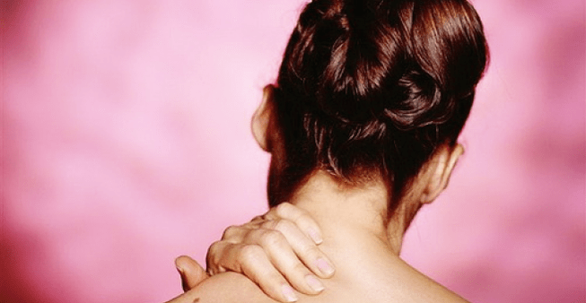
The main symptoms of cervical osteochondrosis
At the level of the cervical segments, the nerve roots and their arteries, the spinal cord and their spinal vessels and arteries can be subjected to compression.Compression of the spinal cord is possible due to posterior intervertebral hernia or rear osteophytes.People with a narrow vertebral channel are especially predisposed to this.With a hernia, the signs of cervical osteochondrosis compression develop rapidly and the symptoms of the cephalorraquidian fluvin current block are softer.
It is very difficult to clinically distinguish the compression of the spinal cord with a tumor and hernia.Cervical spine osteochondrosis is manifested by a spastic paresis of the legs, conduction disorders of sensitivity, pain and weakness in the hands.In some cases, signs of compression are combined with signs of spinal cord ischemia that emerged as a result of spinal artery compression and root vessels.
Symptoms of damage to previous horns and ventral departments can suddenly develop with the involvement of pyramidal paths (blood supply to anterior spinal artery).Anterior spine syndrome occurs: slow paresis of the arms, spastic paresis of the legs, impaired sphincter function.Sometimes the symptoms of gross violation of profound sensitivity in the hands develop.After 2-3 weeks, the signs of a blow to the spine begin to regress.In terms of the volume of pathological focus, we can say about the severity of residual phenomena.
Cervical myelopathy
Myelopathy is a chronic ischemization for cervical osteochondrosis.A major role in the development of this syndrome is played by compression of blood vessels.The most characteristic is the defeat of the ventral parts of the side pillars and the front horns.It is manifested by a sposticrootic paresis of the arms, a spastic paresis of the legs, a violation of the profound sensitivity of the legs (classic triad).
In several patients, the symptom of Lermitta appears: a feeling of passing the electrical discharge throughout the spine with irradiation of pain in the hands and legs when heading.It is possible to develop lateral amyotrophic sclerosis in which there are no symptoms of bulbar.
An important role in the confirmation of myolopathy is played by magnetic resonance imaging and CT, which reveal the compression of the peel bag with osteophytes and a thick yellow group.
Signs of root compression
Since underlying discs wear faster, spondylartrrosis develops in the corresponding segments.Osteophytes narrow the intervertebral holes and squeeze the roots (at the lumbar level most often a herniated disc compression in the epidural space).By moving the growth head, the spine is injured, which causes edema formation, which further restricts the intervertebral hole.Develop reactive inflammatory reactions.
Clinical manifestations:
- C3 -koreshok (below 2 cervical vertebra occurs quite rarely) - pain in the corresponding half of the neck, a feeling of swelling of the tongue, a feeling of coma in the throat;
- C4 -KoresShok - Pain in appropriate shoulder flow, clavicle, trapezoid muscle atrophy, a decrease in neck muscles (3 and 4 cervical roots increases the tone of the diaphragm, which leads to a change in the liver and the appearance of the sparked angina);
- C5 -Decor - Pain in the neck and external surface of the shoulder, hypotrophy of the deltoid muscle;
- C6 -Koreshok (one of the most common locations) -Pulle in the neck, blades, shoulder, the radial surface of the forearm spreads to 1 finger, seemed in the hands, weakness of the two tooth muscle biceps;
- C7-Koreshok-Pain spreads to 2-3 fingers, accompanied by paresthesia, three-head muscle weakness;
- C8 -Koreshok - The pain extends to the surface of the forearm elbow to the 5th finger, accompanied by paresthesia.
Cervical reflex syndromes
Vertebral syndrome is manifested by acute cervical (bastard, cervical) pain, less often chronic or subacute pain.The main sources of pain syndrome are a fibrous ring, the rear longitudinal ligament, joint capsule, tense muscles.Krivosheya is not as pronounced as the curvature of the spine at the lumbar level.
The pains are sore, radiate to the back of the head.Intensify when driving or prolonged in a position.In palpation, it is determined the pain of the processes and thorny capsules of the joints on the painful side (along the posterior surface of the 3-4 cm neck is lateral that spicy processes) is determined.Involvement in the process not only of the back, but also of the frontal spine muscles (anterior ladder, etc.) is characteristic.
Anterior staircase syndrome
The muscle tension of the ladder occurs very often with cervical osteochondrosis.The muscle is determined by the side of the sternum -shaped muscle in the form of stressful, dense and increased size compared to the healthy side.Due to tension, the compression of the supravific vessels, which is accompanied by pain and swelling in the hand, impaired sensitivity and motor activity (along the elbow nerve).The pain intensifies in a horizontal position.
Small Pesestic Muscle Syndrome
The development mechanism is similar to the previous one.Compression of the vascular freezing beam occurs between the muscle and the shoulder bone (or heart process) in conditions of increased hand abduction.It is accompanied by chest pain, shoulder, hand.
Existing characteristics are often considered as heart pain with VSD (there are no acute attacks, the effect of taking nitroglycerin or sedatives is not increased symptoms during movement and palpation of problem points).
The rear friendly syndrome
Distable and vasomotor disorders that occur as a result of the irritation of the sympathetic plexus of the vertebral artery are characteristic.Plexus branches are located in brain and skull tissues.It is clinically manifested by dizziness, a touch of ears, spectacular disorders, anxiety.
Compression of vertebral arteries with osteophytes emanating from the articulations of the spine spine, in combination with atherosclerotic damage to these vessels, is an important pathogenetic factor in the development of brain and spinal cord insufficiency.
Conclusion
In most cases, hand and neck pain is associated with cervical osteochondrosis.In some patients, pain is caused by the hernia of the intervertebral disc in others - osteophytes and arthrosis of the spine joints.Each of these options can lead to local or reflected pain, root syndrome and myelopathy.When examining patients with neck pain, it is necessary to exclude pathologies such as:
- spine tumors;
- epidural abscess;
- Spondylitis;
- subarachnoid hemorrhage;
- meningitis;
- Salon abscess;
- stratification of carotid artery;
- Cervical vertebra fracture.